Medical imaging routinely used in cancer care may hold far more biological information than previously thought, according to an international study involving Umeå University guest professor Lukas Kenner. The research shows that PET/CT scans can capture the molecular activity of aggressive head and neck tumors, potentially improving precision diagnosis.
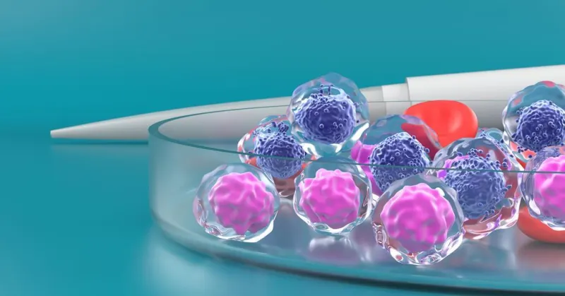
The study, published in a peer-reviewed journal, analyzed PET/CT scans from patients with head and neck squamous cell carcinoma. By examining the metabolic and molecular signatures captured by the imaging, researchers found that these scans could differentiate between tumor subtypes with varying aggressiveness. This could help clinicians tailor treatments more effectively.
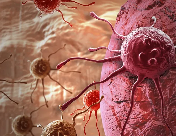
Professor Kenner, who is also affiliated with the Medical University of Vienna, emphasized that the findings could lead to non-invasive biopsies. Instead of relying solely on tissue samples, doctors might use imaging data to assess tumor biology in real time. The approach could reduce the need for repeated invasive procedures.
The research team plans to validate the results in larger clinical trials. If confirmed, the technique could become a standard tool for managing head and neck cancers, which account for about 4% of all cancers globally. Early and accurate diagnosis is critical, as these tumors often spread quickly.